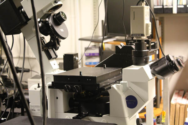
Total Internal Reflection Fluorescence Microscope
This microscope uses objective type total internal reflection excitation at the top surface of the lower microscope coverslip. The fluorescence is detected with a back illuminated on-chip multiplication gain charge-coupled-device (CCD) camera. The image is split into two channels (donor and acceptor fluorophores if doing FRET, orthogonal polarizations if doing anisotropy). The wide field configuration is well suited for simultaneous monitoring of the fluorescence of many single molecule (i.e., protein/nucleic acid complexes) undergoing irreversible chemical reactions. This setup allows monitoring a large (70 μm x 70 μm) region, ca. 250 single molecules can then be simultaneously observed, albeit with a lower time resolution than in the confocal configuration (ca. 15 ms). The setup further allows transmission measurements in DIC mode. The setup consists of an inverted fluorescence microscope (Olympus IX-71) in conjunction with a back-illuminated electron multiplying charge coupled device (EM-CCD) detector (Cascade II:512; Roper Scientific, Tucson, AZ). Excitation sources consist of a diode-pumped solid-state green laser (532-nm cw from Crystal Laser) a diode-pumped solid-state red laser (638 nm cw from Crystal Laser) and an Ar+ laser (outputs at 458 nm, 488 nm, and 514 nm). The laser beam is focused onto the back focal plane of the objective using a TIRF illumination module (IX2-RFAEVA-2, Olympus) to yield an evanescent wave excitation at the glass-water interface. The camera is controlled using Image-Pro Plus 5.1 (Media Cybernetics), capturing 8-bit 512 x 512 pixel images with a conversion gain of 3, and multiplication gain of 4095. Image processing is performed either with Image-Pro Plus or with a self-written algorithm in IDL and Matlab.